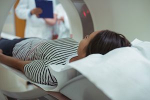
Dr. Jose Pizarro Explores the Uses of Radiology to Diagnose Many Injuries and Diseases
Most patients are familiar with the basic job functions of a radiologist. For example, if a child falls, an X-ray will be taken to determine whether the child has broken a bone. This technology has existed for over a hundred years and has developed into an exciting field full of many essential advancements.
Dr. Jose Pizzaro, a respected neuroradiologist from Longboat Key, Florida, explains how radiology goes beyond broken bones and diagnoses problems in many areas of the body. The course of scientific innovations is tracked for several locations within the human body.
Radiology of the Brain

One of the most important applications of radiological science is in scanning the brain. Until recently, the brain could not effectively be studied in a living person. Advances in radiological scans have enabled doctors to effectively diagnose strokes, brain tumors, circulation problems, and neuropsychiatric conditions like Alzheimer’s disease.
On the brain, CT (computed tomography) and MRI (magnetic resonance imaging) are often used together to diagnose various ailments. These scans can detect blood flow constrictions and obstructions that cause stroke, and they can help doctors determine whether it is safe to operate on a brain tumor.
Transcranial Doppler ultrasound measures blood flow to the large arteries of the brain. It is used to detect blockages and other conditions that increase the risk of stroke.
Brain scans have also been used to detect problems that stem from severe COVID-19 infections. Problems with encephalopathy, seizures, cerebellar systems, and peripheral nervous system disorders were observed. Sixty-eight percent of the patients in this study had abnormal brain pathologies.
One of the newest techniques using radiology to study the brain is called diffusion tensor imaging. With this technique, the flow of fluid within the brain can be tracked, leaving doctors with images of where gray and white matter is located. The technique involves taking several MRI images that are calibrated to view the flow of water in the brain between different types of tissue.
Using diffusion tensor imaging means that doctors can better judge the safest routes for removing lesions like gliomas and other brain tumors.
Another use of diffusion tensor imaging is creating an image of the brain’s gray matter. This technique is known as fiber tractography. This enables neuroradiologists to create a 3-D map of the brain showing all of its vulnerable areas.
The Nervous System
The spinal canal can be studied using myelography, which is an X-ray study. By injecting an iodine-based dye into the spinal fluid, the structures can be seen in X-ray images. This procedure can diagnose blockages in the spinal canal caused by arthritis, herniated discs, infections, or tumors.
Cardiac Applications
Radiology of the heart has undergone many exciting developments over the past several years. The heart can be studied using ultrasound, EKG testing, ECG testing, and MRI, among other techniques. Cardiac catheterization is used to determine how much damage a patient’s heart experienced during an adverse event such as a heart attack. Patients are examined through catheterization to find whether they may have further blockages or abnormalities in their heart that could cause more problems. During the catheterization process, doctors can also perform interventional therapies like inserting stents.
Reproductive Systems
Ultrasound is commonly used for both the male and female reproductive systems. Systemic abnormalities can be diagnosed, and the health of a pregnancy can be checked. A female patient’s fallopian tubes can be examined for blockages that may be causing infertility in a hysterosalpingogram.
Bones and Muscles
Radiology can also be used to diagnose problems with bones and muscles. In addition to X-rays, the traditional method of examining bones, radiologists can use MRI and CT scans. An X-ray is a relatively simple and cost-effective method of getting images, but MRI and CT scans have their advantages.
MRI scans can be used to examine the bones and joints and soft tissues like tendons, muscles, and cartilage. These scans are beneficial for checking for structural abnormalities, tumors, bone marrow disease, and herniation of spinal cord discs. They are also frequently used for patients with arthritis. These scans can help doctors understand how their patients’ conditions have progressed.
The Impact of Radiology
Radiology can provide effective means of helping doctors to understand how the body works and how certain conditions may be affecting it. The science of radiology has developed a great deal since its first uses on the battlefield in the late 19th century. Patients have come to benefit a great deal from applying these technologies.
Having a good grasp of how radiology affects the medical field can help a person understand how much medical science has evolved over the past decades. Radiologists and neuroradiologists like Jose Pizzaro have been hard at work advancing their craft.
Dr. Jose Pizzaro believes that the science of radiology will continue to evolve a great deal in the coming years, introducing techniques that will help patients recover more easily from conditions affecting all systems in the body.


